Scientists at the Wake Forest Institute for Regenerative Medicine have developed a new 3D printer able to accurately produce models of human body parts. The group of scientists has successfully transplanted living tissue created by the machine, according to a study published in the journal Nature.
Using a combination of living cells and a special gel, researchers have been able to print out human body parts such as muscles, jawbones and even full-grown ears.
The research represents a groundbreaking advance in regenerative medicine, as it shows the possibilities for tissues to be successfully transplanted into patients. However, researchers at the Wake Forest University noted a lot of technical obstacles holding back the process.

Although the current tissue transplant survived when implanted into animals, there’s still research to be done before human transplants can be performed.
“We show that we can grow muscle. We make ears the size of baby ears,” Dr. Atala said to NBC News. “We make jawbones the size of human jawbones. We are printing all kinds of things,” he added.
The group of scientist accountable for the study, led by Dr. Anthony Atala named the bioprinting process as ‘the integrated tissue and organ printing system’ or ITOP.
The use of a 3D printed organ goes beyond the transplant because it can be the object of multiple tests. Previous attempts showed badly printed out organ shapes as they were not solid and ended up dying.
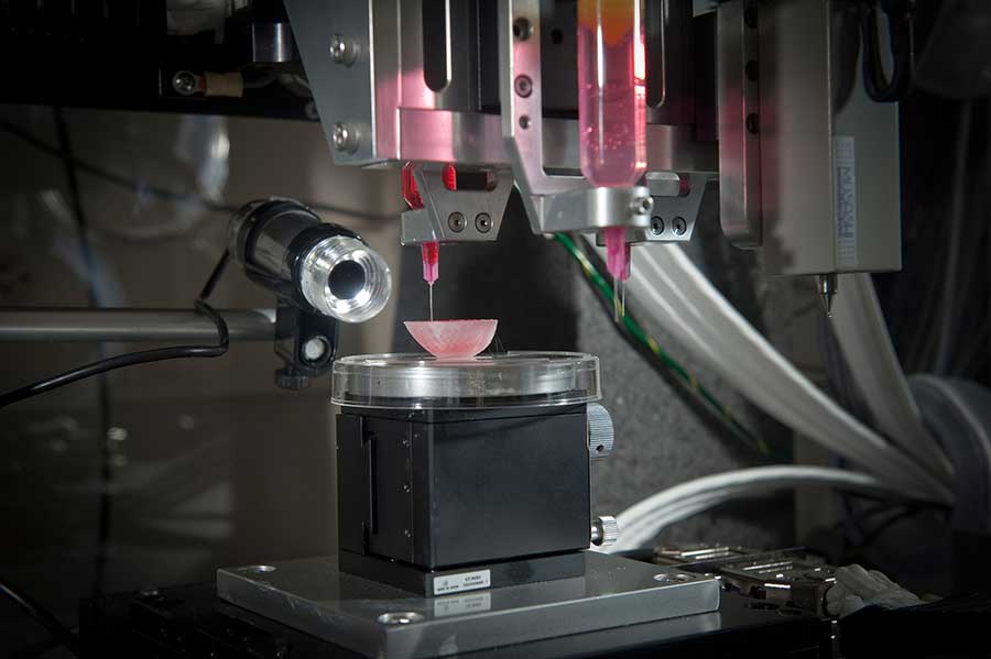
Mechanical machines creating live body parts.
The new custom bioprinter creates the implants by layering patterns of a mixture of cells and a biodegradable gel. This new approach solved the implant’s adaptability by creating organs with tiny tunnels within them.
This allows tunnels to serve as a passage for nutrients until blood vessels can grow naturally, the study suggests. Wake Forest scientists were able to print stable cartilage, bone and muscle structures and successfully transplanting them into rodents.
The new device capable of printing body parts is programmed to slowly print layer upon layer of a rapidly hardening material in the form of droplets. The process, however, is pretty much like previous 3D printers used to create objects, by printing complex shapes in three dimensions accurately.
Still, the key to the machine’s success is the combination of living cells with a biodegradable plastic called polycaprolactone. Serving as a solid structure, it supports the organ while it’s being fabricated and the growing cells take root, and then it falls apart.

No army left behind
The study led by Dr. Atala was made possible thanks to funding. The research received funding from the Armed Forces Institute of Regenerative Medicine, a federally funded organization.
The purpose of which is to apply any consistent findings to those injured on the battlefield. The use of regenerative medicine is a crucial fact for the federal organizations as it provides safety measures never seen before. If wounded on the battlefield, the army could fabricate the body parts needed for surgery on demand, instead of waiting for an organ donor.
However, the US Army Research Office is already using the normal 3D printer, as they look for alternative combat armor options. New body armors are being developed by the MIT to maximize the ability to move while providing more protection than Kevlar body armors.
So, whether they can produce living organs or alternative military applications 3D printing and bioprinting are surely under the army’s sight.
Source: The Verge