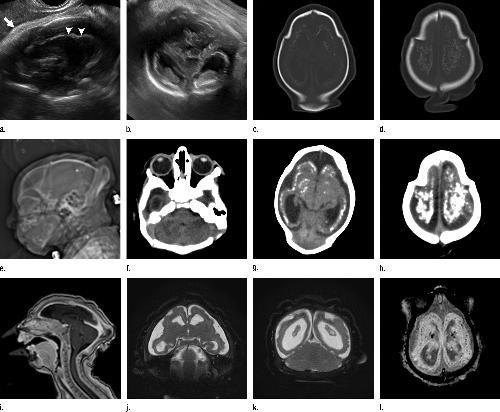
The more health experts learn from the Zika virus, the more macabre it becomes. A study published Tuesday in the journal Radiology revealed a shocking collection of brain scans and ultrasound pictures showing the devastation that 45 Brazilian babies suffered because their mothers were infected with Zika during pregnancy.
The researchers found that the virus’ threats go beyond microcephaly and that the first semester is the time where infections are riskiest for the pregnancy. Study coauthor Dr. Deborah Levine, head of Obstetric & Gynecologic Ultrasound at Beth Israel Deaconess Medical Center, affirmed that the abnormalities in the fetal brain caused by the Zika virus are very severe compared to other congenital infections, according to a report by Fox News.
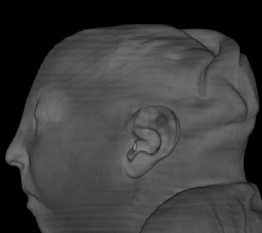
The research team was surprised to learn that the virus was found to be linked not only to microcephaly, a condition known for causing extremely small heads in newborns as a result of an underdeveloped brain, but also to share a strong connection with eye defects, stunted growth and hearing impairment.
Zika’s impact on the babies’ brain
The study authors demonstrated in their report that fetuses exposed to the virus were also at risk of suffering from gray and white matter volume loss, brainstem abnormalities, and calcifications. Another brain abnormality the researchers found was ventriculomegaly, a condition that consists of enlarged ventricles.
The majority of the babies involved in the study were born with microcephaly, but some of them suffered from damages of the corpus callosum, a crucial part of the brain in charge of connecting the two hemispheres.
“It’s not just the small brain, it’s that there’s a lot more damage,” said Dr. Levine, according to The New York Times. “The abnormalities that we see in the brain suggest a very early disruption of the brain development process.”
The virus was found to have played a major role in the damage of the cerebellum, which is linked to speech, balance, and movement. Moreover, the basal ganglia, involved in the processes of thinking and emotion, was included in the brain parts affected by the Zika virus.
The images observed in the study came from 28 babies with presumed exposure to the virus and 17 whose mothers had a confirmed infection during pregnancy.
Dr. Levine and his colleagues analyzed pictures from the Instituto de Pesquisa in Paraiba, located in the northeastern part of Brazil. Three of the newborn involved in the research passed away within the first three days of life and study authors conducted studies on their autopsy reports.
The researchers decided to make many of the images public because they wanted doctors around the world to have a better understanding of the threat the virus poses to fetal brains and have a better idea of what to look for in those brains affected by the Zika virus, as The New York Times reported.
Source: The New York Times